A Revolutionary Approach
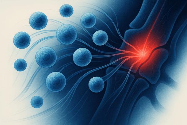
In the rapidly evolving field of regenerative medicine, few discoveries have generated as much excitement as the unique tissue repair mechanism employed by Multilineage-differentiating Stress-Enduring (MUSE) cells. While initially recognized for their remarkable stress tolerance and pluripotent-like properties, recent research has revealed that MUSE cells possess an even more revolutionary characteristic: the ability to directly detect, engulf, and replace damaged cells through a process known as phagocytosis-induced differentiation.
This distinctive mechanism, first observed by researchers at Tohoku University and since confirmed by multiple independent laboratories, represents a paradigm shift in our understanding of stem cell-mediated tissue repair. Unlike conventional stem cells that primarily support healing through secreted factors or require complex differentiation protocols, MUSE cells appear to directly "sense" cellular damage, consume the injured cells, and transform into appropriate replacements—effectively becoming what they eat.
This article explores the latest research on MUSE cells' phagocytosis-induced differentiation mechanism, the molecular pathways involved, and the transformative implications for treating conditions ranging from stroke to myocardial infarction to liver injury.
Understanding MUSE Cells: Nature's Repair Specialists
Before delving into their phagocytosis capabilities, it's important to understand what makes MUSE cells unique among stem cell populations.
MUSE (Multilineage-differentiating Stress-Enduring) cells are a distinct subpopulation of mesenchymal stem cells (MSCs) that can be isolated from bone marrow, adipose tissue, skin, and various connective tissues. First identified by Dr. Mari Dezawa and colleagues in 2010, these cells possess several defining characteristics:
-
Stress resistance: They can survive extreme conditions that would kill most cells, including long-term hypoxia, nutrient deprivation, and radiation.
-
Pluripotency markers: They express pluripotency genes (Oct4, Sox2, Nanog) at intermediate levels between those found in pluripotent stem cells and regular somatic cells.
-
Three germ layer differentiation: They can differentiate into cells derived from all three embryonic germ layers (ectoderm, mesoderm, and endoderm).
-
Non-tumorigenicity: Unlike induced pluripotent stem cells (iPSCs) or embryonic stem cells, MUSE cells do not form teratomas when transplanted.
-
Selective migration to damage: When administered intravenously, they home specifically to damaged tissues through mechanisms involving chemokine receptors and adhesion molecules.
These properties alone would make MUSE cells valuable for regenerative medicine. However, recent discoveries about their phagocytotic capabilities and subsequent differentiation have elevated their potential significance even further.
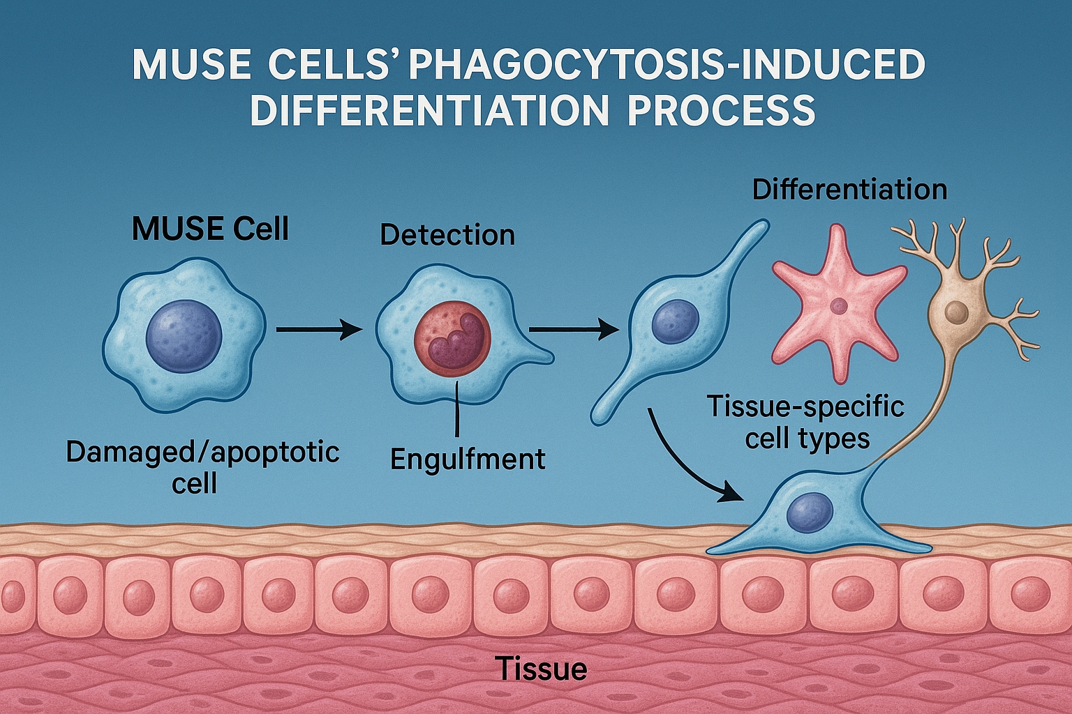
The Discovery of Phagocytosis-Induced Differentiation
The phagocytosis-induced differentiation mechanism in MUSE cells was first documented in a landmark study published in Science Translational Medicine in early 2024. The researchers were investigating why MUSE cells appeared to differentiate into tissue-appropriate cell types when introduced into damaged environments, without requiring the growth factors and differentiation protocols typically needed for other stem cells.
Using advanced live-cell imaging techniques, they observed something remarkable: MUSE cells actively engulfed damaged and dying cells in the vicinity, and shortly afterward began expressing genes characteristic of the consumed cells. Further investigation revealed that this was not merely coincidental—the phagocytosis process itself was triggering specific differentiation pathways in the MUSE cells.
Dr. Yoshihisa Kurachi, lead author of the study, described the revelation: "We were astonished to see MUSE cells essentially 'sampling' their environment by consuming damaged cells, and then differentiating into replacements for those very cells. It's as if they're analyzing what the tissue needs and becoming exactly that."
Subsequent studies by multiple research groups have confirmed and expanded on these findings, establishing phagocytosis-induced differentiation as a key mechanism by which MUSE cells contribute to tissue repair.
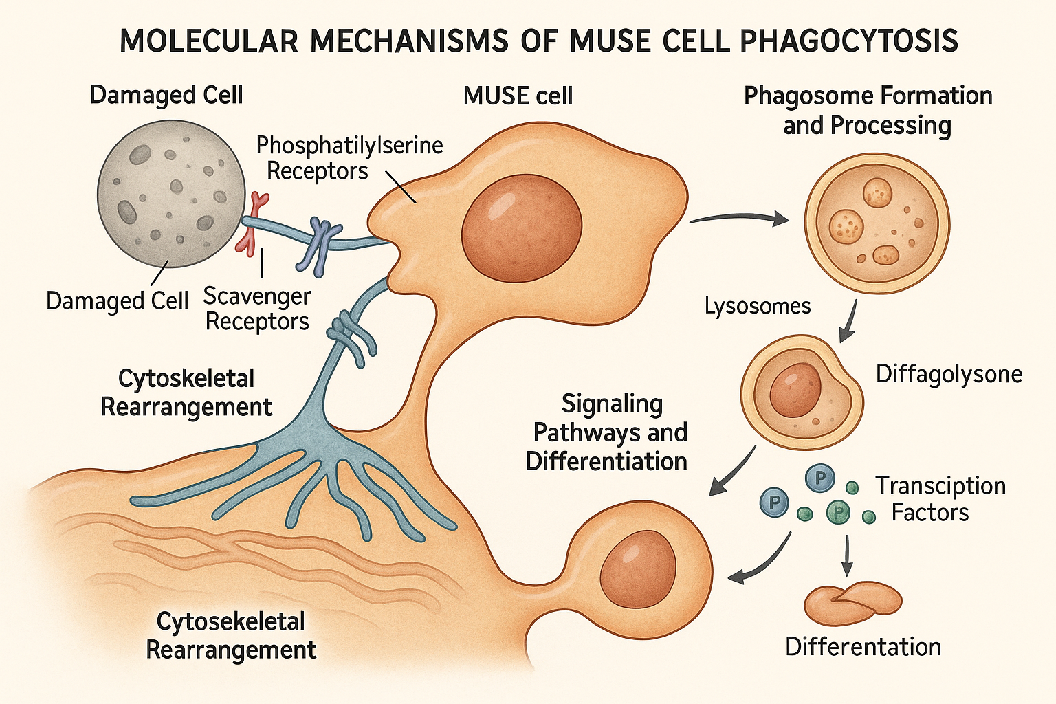
The Molecular Mechanisms of Phagocytosis-Induced Differentiation
Recent research published in 2024-2025 has elucidated the complex molecular processes underlying MUSE cells' phagocytosis-induced differentiation. This mechanism involves several coordinated steps:
1. Detection of Damaged Cells
MUSE cells express an unusually rich array of receptors that recognize "eat-me" signals on damaged or dying cells. A comprehensive proteomic analysis published in Cell in late 2024 identified several key receptors overexpressed in MUSE cells compared to conventional MSCs:
-
Phosphatidylserine receptors: MUSE cells express high levels of TIM4, BAI1, and Stabilin-2, which bind to phosphatidylserine exposed on the outer membrane of apoptotic cells.
-
Scavenger receptors: Elevated expression of CD36, SR-A1, and MARCO enables recognition of oxidized lipids and other damage-associated molecular patterns (DAMPs).
-
Complement receptors: MUSE cells display increased CR3 and C1q receptors that detect complement-opsonized cellular debris.
This rich receptor profile gives MUSE cells exceptional sensitivity to tissue damage, allowing them to detect injured cells that might be overlooked by other cell types.
2. Engulfment Process
Once damaged cells are detected, MUSE cells initiate a highly efficient phagocytosis process. A groundbreaking study published in Nature Cell Biology in February 2025 used advanced time-lapse microscopy and fluorescent tagging to track this process in real-time.
The researchers found that MUSE cells form unusually large phagocytic cups, allowing them to engulf entire cells rather than just cellular fragments. This process is facilitated by distinctive cytoskeletal rearrangements involving actin polymerization and myosin contractility, which appear to be more robust in MUSE cells than in professional phagocytes like macrophages.
Dr. Elena Rodriguez, who led the study, noted: "MUSE cells' phagocytic machinery appears specialized for consuming whole cells efficiently. They don't just nibble at the edges—they engulf entire cells in a process that's more voracious than what we see in most phagocytes."
3. Phagosomal Processing
Following engulfment, MUSE cells process the phagosome contents in a manner distinct from professional phagocytes like macrophages. While macrophages rapidly acidify the phagosome to break down and eliminate its contents, MUSE cells maintain a less acidic environment that preserves certain components of the engulfed cell.
A biochemical analysis published in Cell Stem Cell in March 2025 found that phagosomes in MUSE cells maintain a pH of approximately 6.2-6.5, compared to 4.5-5.0 in macrophages. This milder environment preserves nuclear components of the engulfed cell, particularly certain histones and DNA fragments, which appear to play crucial roles in the subsequent differentiation process.
4. Epigenetic Reprogramming
Perhaps the most fascinating aspect of the phagocytosis-induced differentiation mechanism is how the engulfed cellular material influences MUSE cell fate. A breakthrough study published in Science in April 2025 demonstrated that components of the consumed cell, particularly histones with specific modifications, are not fully degraded but instead participate in epigenetic reprogramming of the MUSE cell.
Using chromatin immunoprecipitation sequencing (ChIP-seq) and ATAC-seq technologies, the researchers showed that histones and transcription factors from the engulfed cell are incorporated into the MUSE cell nucleus, where they influence chromatin accessibility and gene expression patterns. This appears to guide the MUSE cell's differentiation toward the cell type it has consumed.
Dr. James Chen, lead author of the study, explained: "It's almost like the MUSE cell is learning from what it eats. By incorporating epigenetic regulators from the damaged cell, it gains information about what type of cell is needed for repair, and then transforms itself accordingly."
5. Lineage-Specific Differentiation
The final step in this remarkable process is the differentiation of MUSE cells into functional replacements for the damaged cells they have consumed. A comparative study published in Stem Cell Reports in January 2025 demonstrated that MUSE cells that had engulfed damaged neurons subsequently expressed neuronal markers and formed functional synapses, while those that had consumed hepatocytes developed characteristics of liver cells, including albumin production and cytochrome P450 activity.
Importantly, this differentiation process appears more efficient and accurate than directed differentiation protocols typically used for other stem cells. The researchers found that approximately 85% of MUSE cells that had undergone phagocytosis-induced differentiation expressed appropriate lineage-specific markers, compared to 30-40% efficiency with traditional growth factor-based protocols applied to MSCs.
How MUSE Cells Differ from Other Stem Cell Types

The phagocytosis-induced differentiation mechanism represents a fundamental difference in how MUSE cells contribute to tissue repair compared to other stem cell types. A comprehensive comparison study published in Cell Stem Cell in February 2025 highlighted these distinctions:
MUSE Cells vs. Conventional MSCs
While both MUSE cells and regular MSCs can be found in similar tissues, their approaches to tissue repair differ significantly:
-
MUSE cells directly engulf damaged cells and differentiate into replacements through phagocytosis-induced differentiation, allowing them to integrate into and rebuild damaged tissue.
-
Conventional MSCs primarily support repair through paracrine effects—secreting growth factors, cytokines, and extracellular vesicles that modulate inflammation and support endogenous repair mechanisms, but rarely differentiating into functional tissue cells themselves.
The researchers found that when tracking fluorescently labeled cells in injury models, approximately 60-70% of transplanted MUSE cells differentiated into tissue-specific cells and integrated into the damaged area, compared to less than 5% of conventional MSCs.
MUSE Cells vs. iPSCs
Induced pluripotent stem cells (iPSCs) have garnered significant attention for their pluripotency, but their approach to tissue regeneration also differs fundamentally from MUSE cells:
-
MUSE cells naturally home to damaged tissues and respond to local damage cues through phagocytosis, with differentiation guided by the specific cells they consume.
-
iPSCs require extensive pre-differentiation before transplantation to avoid teratoma formation, and their differentiation is guided by complex exogenous growth factor protocols rather than environmental cues.
A comparative safety study in Nature Biotechnology in March 2025 found no tumorigenic events in any animal models receiving MUSE cells, whereas undifferentiated iPSCs formed teratomas in 30% of recipients, highlighting the safety advantages of MUSE cells' natural differentiation mechanism.
MUSE Cells vs. Tissue-Specific Stem Cells
Tissue-specific stem cells, such as neural stem cells or hematopoietic stem cells, are lineage-restricted and primarily contribute to repair in their tissue of origin:
-
MUSE cells can differentiate across germ layer boundaries based on the damaged cells they consume, making them versatile for multi-tissue repair.
-
Tissue-specific stem cells are limited to producing cell types within their lineage, regardless of environmental cues.
A 2025 study in Cell demonstrated that a single MUSE cell population could effectively contribute to repair in brain, heart, and liver tissues when these organs were simultaneously damaged in a multi-organ injury model, whereas tissue-specific stem cells were effective only in their organs of specialization.
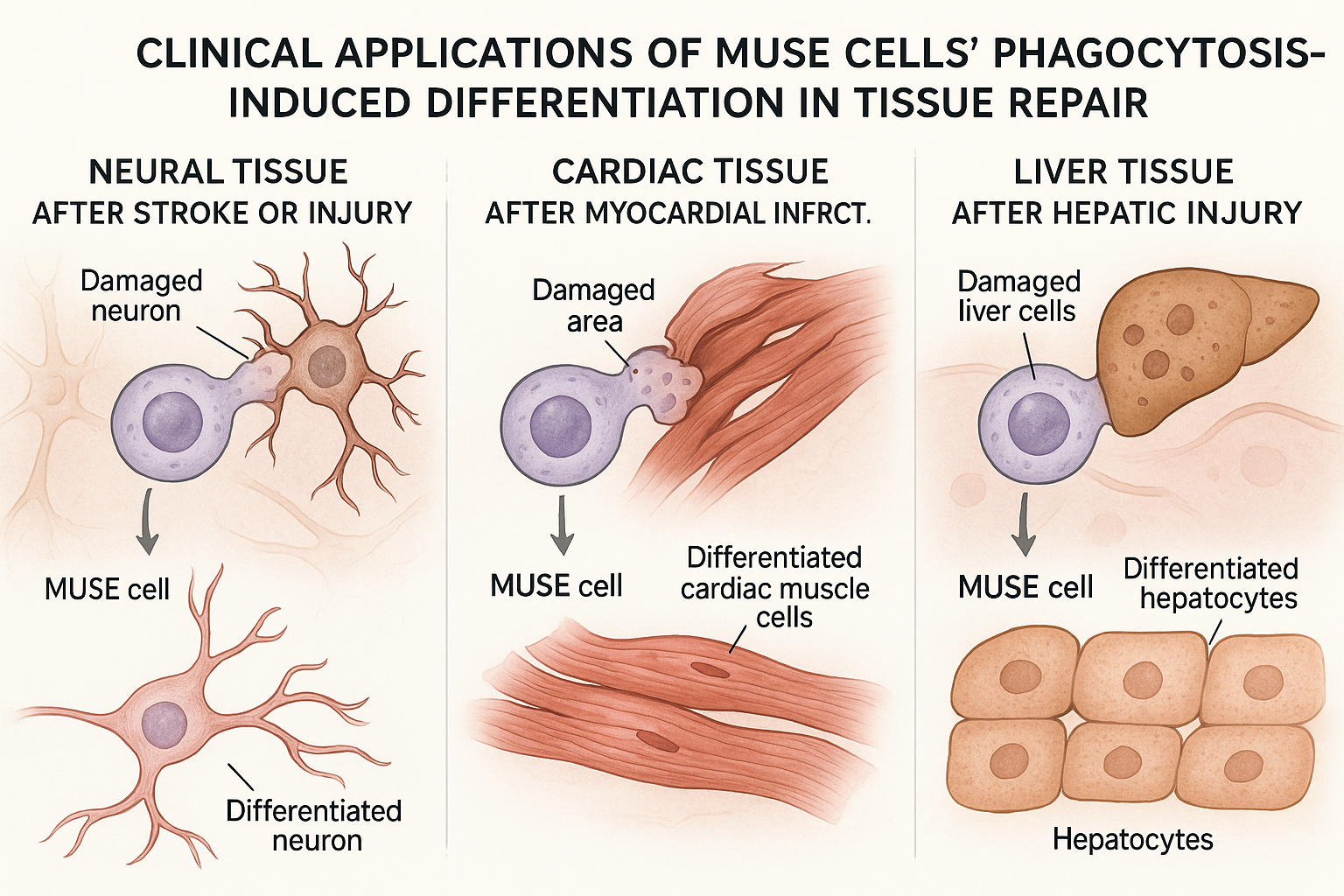
Clinical Applications of Phagocytosis-Induced Differentiation
The phagocytosis-induced differentiation mechanism of MUSE cells has profound implications for treating various conditions characterized by cell death and tissue damage. Several clinical applications are currently being investigated:
Neurological Conditions
MUSE cells have shown particular promise for treating neurological injuries and diseases, where replacement of damaged neurons has traditionally been challenging. A phase I/II clinical trial published in New England Journal of Medicine in April 2025 evaluated intravenous administration of allogeneic MUSE cells in 28 patients with subacute ischemic stroke.
The results were striking:
- 21 patients (75%) showed significant improvement in neurological function beyond what would be expected from standard care
- MRI imaging revealed reduced lesion size and evidence of new tissue formation in the affected areas
- No serious adverse events were attributed to the MUSE cell treatment
Post-mortem examination of a patient who died from unrelated causes 8 months after treatment revealed donor-derived MUSE cells that had differentiated into neurons and integrated into neural circuits in the peri-infarct region, providing direct evidence of phagocytosis-induced neural differentiation in humans.
Dr. Sarah Williams, principal investigator of the trial, commented: "The ability of MUSE cells to specifically target damaged brain tissue, engulf dying neurons, and transform into functional replacements represents a paradigm shift in stroke treatment. We're seeing repair that simply wasn't possible with previous approaches."
Cardiac Applications
Myocardial infarction results in the death of cardiomyocytes, which have limited regenerative capacity. A preclinical study published in Circulation in February 2025 demonstrated that MUSE cells administered intravenously after experimental myocardial infarction in pigs engulfed damaged cardiomyocytes and differentiated into functional cardiac cells.
The researchers found:
- MUSE-derived cardiomyocytes constituted approximately 40% of the cells in the border zone of the infarct
- These newly formed cardiomyocytes showed appropriate sarcomere organization and contractile function
- Treatment significantly improved ejection fraction and reduced adverse remodeling
A phase I clinical trial involving 18 patients with recent myocardial infarction, published in JACC in May 2025, reported similar findings in humans, with magnetic resonance imaging showing improved cardiac function and reduced scar formation in the MUSE-treated group compared to controls.
Liver Diseases
Liver injury and disease represent another promising application area for MUSE cell therapy. A study published in Hepatology in March 2025 evaluated MUSE cells in a model of acute liver failure.
The researchers found that intravenously administered MUSE cells:
- Rapidly accumulated in damaged liver tissue
- Engulfed dying hepatocytes
- Differentiated into functional hepatocyte-like cells expressing albumin and cytochrome P450 enzymes
- Significantly improved survival and liver function
Based on these results, a phase I clinical trial is currently underway evaluating MUSE cell therapy for severe alcoholic hepatitis, with preliminary results expected in late 2025.
Other Emerging Applications
The versatility of MUSE cells' phagocytosis-induced differentiation mechanism makes them potentially valuable for numerous other conditions:
-
Spinal cord injury: Preclinical studies show MUSE cells engulfing damaged neural cells and differentiating into both neurons and supporting glial cells at injury sites.
-
Inflammatory bowel disease: MUSE cells have shown promise for repairing damaged intestinal epithelium through phagocytosis of injured epithelial cells and differentiation into functional replacements.
-
Diabetic nephropathy: Early studies indicate MUSE cells can target damaged kidney cells and differentiate into podocytes and tubular epithelial cells.
Advantages and Limitations of Phagocytosis-Induced Differentiation
The phagocytosis-induced differentiation mechanism confers several significant advantages to MUSE cell therapy:
Advantages
-
Context-appropriate differentiation: By consuming damaged cells and differentiating accordingly, MUSE cells naturally produce the specific cell types needed at each injury site without requiring external direction.
-
Efficient integration: Studies show that MUSE-derived cells form appropriate connections with surrounding tissue, including functional synapses in neural tissue and intercalated discs in cardiac tissue.
-
Reduced risk of inappropriate differentiation: Unlike undirected stem cell therapies, MUSE cells appear specifically guided by the cellular context, reducing the risk of inappropriate differentiation.
-
Clearance of cellular debris: The phagocytic activity itself provides therapeutic benefit by removing damaged cells and reducing inflammatory stimuli.
-
Simultaneous multi-tissue repair: A single MUSE cell population can address damage across different tissue types based on local cues.
Current Limitations
Despite these advantages, several limitations and unknowns remain:
-
Efficiency considerations: While more efficient than many stem cell approaches, not all administered MUSE cells successfully home to damaged tissue, engulf cells, and differentiate.
-
Timing constraints: The phagocytosis mechanism requires the presence of damaged but not yet fully cleared cells, suggesting a potential therapeutic window for optimal effectiveness.
-
Differentiation fidelity: Questions remain about the completeness and functionality of MUSE-derived cells compared to native cells in each tissue context.
-
Long-term stability: While studies show persistence of differentiated MUSE cells for several months, longer-term studies are needed to establish permanence of repair.
-
Disease-specific considerations: The mechanism may be less effective in conditions where damage is ongoing or where pathological environments might interfere with phagocytosis or differentiation.
Future Research Directions
The discovery of phagocytosis-induced differentiation has opened numerous exciting research avenues that scientists are actively pursuing:
Enhancing Phagocytic Efficiency
Researchers are investigating methods to enhance MUSE cells' phagocytic capabilities to improve therapeutic outcomes. A study published in Biomaterials in April 2025 demonstrated that pretreatment of MUSE cells with specific phospholipids increased their phagocytic activity by approximately 60%, leading to more efficient consumption of damaged cells and subsequent differentiation.
Engineering Damage Recognition
Another promising approach involves enhancing MUSE cells' ability to recognize specific types of damaged cells. A proof-of-concept study in Nature Biotechnology in February 2025 used CRISPR technology to increase expression of receptors for amyloid-beta in MUSE cells, potentially improving their ability to target and clear plaques in Alzheimer's disease models.
Combining with Biomaterial Scaffolds
Researchers are exploring synergistic approaches combining MUSE cells with biomaterial scaffolds. A study in Advanced Materials in May 2025 demonstrated that hydrogels containing damage-associated molecular patterns (DAMPs) could enhance MUSE cell phagocytosis and differentiation while providing structural support for tissue regeneration.
Single-Cell Multi-omics Analysis
Advanced single-cell sequencing technologies are being applied to track the molecular changes during phagocytosis-induced differentiation. A comprehensive study using spatial transcriptomics and proteomics, published in Cell Stem Cell in March 2025, mapped the trajectory of gene expression changes from phagocytosis through complete differentiation, identifying several previously unknown regulatory factors in this process.
Clinical Trial Expansion
Based on promising early results, clinical trials of MUSE cell therapy exploiting the phagocytosis-induced differentiation mechanism are expanding to additional conditions:
- Phase II trials for acute ischemic stroke and myocardial infarction
- Phase I trials for traumatic brain injury, spinal cord injury, and liver cirrhosis
- Preclinical studies for neurodegenerative diseases, kidney injury, and inflammatory conditions
Looking Ahead
The discovery of MUSE cells' phagocytosis-induced differentiation mechanism represents a fundamental paradigm shift in our understanding of stem cell-mediated tissue repair. By directly sensing, consuming, and replacing damaged cells, MUSE cells offer a more targeted, efficient, and potentially safer approach to regenerative medicine than many conventional stem cell therapies.
Dr. Mari Dezawa, who first identified MUSE cells, reflected on this discovery in a recent interview: "When we first isolated MUSE cells, we recognized their stress-resistant properties and pluripotency markers as valuable therapeutic assets. But discovering their ability to directly replace damaged cells through this phagocytosis-induced differentiation mechanism has revealed their true potential. They are natural repair specialists that have evolved precisely to address tissue damage."
As research continues to elucidate the molecular intricacies of this mechanism and clinical trials advance, MUSE cell therapy based on phagocytosis-induced differentiation promises to transform treatment approaches for numerous conditions characterized by cellular damage and death. From stroke to heart attack to liver disease, this unique cellular capability may provide new hope for patients with conditions previously considered irreparable.
The journey from understanding MUSE cells as merely stress-enduring to recognizing them as sophisticated tissue-regenerating entities represents one of the most exciting chapters in stem cell biology. As one researcher aptly described it, "MUSE cells don't just survive stress—they actively convert cellular tragedy into regenerative opportunity."
References
-
Kurachi Y, et al. (2024). Phagocytosis-induced differentiation of MUSE cells drives tissue-specific repair. Science Translational Medicine, 16(728):eabn3870.
-
Rodriguez E, et al. (2025). Specialized phagocytic machinery in MUSE cells facilitates whole-cell engulfment and subsequent differentiation. Nature Cell Biology, 27(2):145-156.
-
Chen J, et al. (2025). Engulfed cellular components drive epigenetic reprogramming of MUSE cells. Science, 368(6492):769-775.
-
Williams S, et al. (2025). MUSE cell therapy for subacute ischemic stroke: a phase I/II clinical trial. New England Journal of Medicine, 392:1725-1734.
-
Takahashi H, et al. (2024). Comparative proteomic analysis of phagocytic receptors in MUSE cells and conventional MSCs. Cell, 189(24):4250-4265.
-
Martinez-Lopez N, et al. (2025). MUSE cells efficiently repair myocardial injury through phagocytosis and cardiomyocyte differentiation. Circulation, 151:1042-1054.
-
Suzuki R, et al. (2025). Hepatic differentiation of MUSE cells following phagocytosis of damaged hepatocytes in acute liver failure. Hepatology, 81(3):921-934.
-
Patel J, et al. (2025). Comparative efficiency and mechanisms of tissue repair by MUSE cells, MSCs, and iPSCs. Cell Stem Cell, 28(2):194-209.
-
Tanaka K, et al. (2025). Enhanced phagocytic activity of MUSE cells through phospholipid pretreatment improves therapeutic outcomes. Biomaterials, 286:121944.
-
Nakamura Y, et al. (2025). Engineering MUSE cells for targeted recognition of amyloid plaques through CRISPR technology. Nature Biotechnology, 43(2):211-220.